SERVICES
General X-Ray
We pride ourselves on delivering top quality diagnostic imaging services in a timely manner. Using the latest in X-Ray technology, our experienced Radiologists interpret images and provide reports in a completely filmless environment.
Ultrasound
Our dedicated team of Radiologists and Sonographers are available to ensure that your procedures are conducted quickly and professionally. Our state of the art ultrasound machines are completely digital. At all times, we will make every effort to accommodate your needs as quickly as possible.


Mammography
Advanced 3D Tomosynthesis Mammography
We are proud to introduce advanced 3D Tomosynthesis Mammography as part of our commitment to providing the latest in breast screening technology.
What is 3D Tomosynthesis Mammography?
3D Tomosynthesis delivers a new standard in breast imaging. Instead of flat pictures, this tech produces a series of thin layered images, essentially forming a “3D mammogram.” During the scan, an X-ray arm moves slightly over the breast, capturing detailed images in moments. The X-ray dose is akin to a traditional mammogram. A computer then builds a layered 3D picture of the breast tissue.
This advancement lets our radiologists observe breast tissue with unmatched accuracy. Previously hidden details due to tissue overlap now emerge with striking clarity.
iCAD: Advanced Artificial Intelligence for Enhanced Detection
Beyond 3D imaging, we incorporate the AI tech, iCAD, elevating our screening precision. iCAD acts as an additional layer of expert review, aiding in spotting irregularities and potential cancer signs. Grounded in rigorous AI research, clinical tests prove that iCAD enhances cancer detection without extra procedures.
Our commitment is ensuring top breast health care. With 3D Tomosynthesis and iCAD integration, we prioritize your health and peace of mind.
We are accredited by CAR, the Canadian Association of Radiologists, and proud to be affiliated with Alberta Health Services’ Breast Cancer Screening Program (ABCSP).

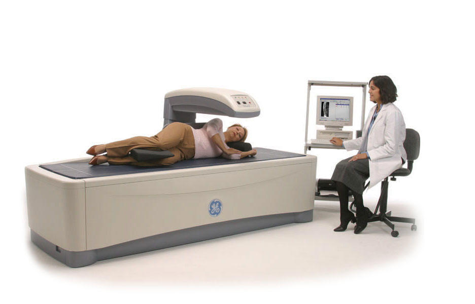
Bone Densitometry
Using only the best DEXA equipment, our team of medical professionals is dedicated to providing you with accurate testing without lengthy waiting lists.
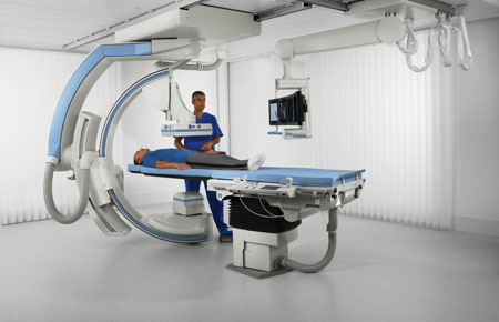
Fluoroscopy / Upper GI
Upper Gastrointestinal (GI) studies utilize the medical imaging technique fluoroscopy, which use x-ray technology to produce real time images of an internal body structure. For GI Studies, fluoroscopy produces images that provide detailed information to physicians to assist in treating or diagnosing various stomach and intestinal illnesses or conditions.
Pain Management / Injections
Fluoroscopy is a medical imaging technique that uses x-ray technology to produce real time images of an internal body structure. Injected medication (usually cortisone) is guided to the area of pain (usually a joint) by fluoroscopy. This allows for a visual on the placement and position of the needle and effective delivery of the medication to the area causing pain.
OUR PROCESS
WE CARE ABOUT YOU
Minimal wait times with less than 24 hr wait time for booked appointments.
MEDICAL ADVICE
Timely reports (8 hour turnaround!) and sate-of-the-art Diagnostic Imaging.
EASY ACCESS
Exceptional customer service. Excellent access and convenient location. Ample free parking and wheelchair accessibility